What results from increasing the packet length when using color Doppler imaging?
A. Decreased penetration
B. Increased aliasing
C. Increased color noise
D. Decreased frame rate
Explanation: Increasing the packet length in color Doppler imaging means increasing the number of pulses used to interrogate each scan line. This improves the accuracy of velocity measurements and sensitivity to low flow velocities but has the drawback of decreasing the frame rate. A longer packet length requires more time to acquire the necessary data, which reduces the number of frames that can be processed and displayed per second. Consequently, while color Doppler imaging becomes more precise, the temporal resolution (frame rate) decreases.
Which change can be made in order to avoid exceeding the Nyquist limit?
A. Increase output power
B. Decrease output power
C. Increase pulse repetition frequency
D. Decrease pulse repetition frequency
Explanation: To avoid exceeding the Nyquist limit and prevent aliasing in Doppler ultrasound, the pulse repetition frequency (PRF) should be increased. The Nyquist limit is half of the PRF, so by increasing the PRF, the Nyquist limit is raised, allowing the system to accurately measure higher velocities without encountering aliasing artifacts.
What is an advantage of power Doppler over color Doppler?
A. Accurate velocity information
B. Increased frame rate
C. Diminished flash artifact
D. Less angle dependent
Explanation: Power Doppler, unlike color Doppler, is less angle dependent because it detects the strength of the Doppler signal rather than the velocity of the blood flow. This means it is more sensitive to detecting low-velocity flow and flow in smaller vessels, regardless of the angle between the ultrasound beam and the flow direction. Color Doppler provides information on flow direction and velocity but is highly dependent on the angle of insonation, making it less reliable when the angle is suboptimal.
Which will affect the gray-scale of a 2-D image?
A. Doppler gain
B. Depth of field
C. Pulse repetition frequency (PRF)
D. Dynamic range
Explanation:
What is the effect of an increased aperture in a linear array transducer?
A. Deeper focus
B. Improved axial resolution
C. Shorter near-field length
D. Decreased temporal resolution
Explanation: The aperture of a transducer is the active area that emits and receives the ultrasound waves. In a linear array transducer, increasing the aperture (using more elements for transmission and reception) results in a deeper focus because the beam is more tightly focused over a longer distance. This improves lateral resolution at greater depths, as the ultrasound beam maintains a narrower width for a longer distance. It allows for better imaging of deeper structures without sacrificing resolution.
What is the primary factor that improves lateral resolution?
A. Propagation speed
B. Beamwidth
C. Frequency
D. Frame rate
Explanation: Lateral resolution refers to the ability of the ultrasound system to distinguish two structures that are side by side, perpendicular to the direction of the sound beam. This resolution is primarily improved by reducing the beamwidth. A narrower beamwidth allows for better differentiation between adjacent structures, enhancing the lateral resolution. Higher frequency transducers can also help achieve a narrower beamwidth, but beamwidth is the primary factor.
Which of these modes has the highest duty factor?
A. Continuous wave Doppler
B. Pulsed wave Doppler
C. Color flow Doppler
D. Gray-scale
Explanation: The duty factor is the fraction of time that the ultrasound system is actively transmitting a signal. Continuous wave (CW) Doppler has the highest duty factor because it continuously transmits and receives ultrasound waves. Unlike pulsed wave Doppler, which alternates between sending and receiving signals, CW Doppler does not have a listening period, resulting in a duty factor of nearly 100%. Therefore, CW Doppler has the highest duty factor among the modes listed.
Which control should a sonographer use to change contrast resolution?
A. Reject
B. Output power
C. Gain
D. Dynamic range
Explanation:
In this image, which artifact is demonstrated?
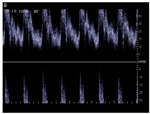
A. Mirroring
B. Aliasing
C. Range ambiguity
D. Spectral broadening
Explanation: The artifact demonstrated in the image is mirroring. This occurs when the ultrasound beam encounters a strong reflector, such as a diaphragm or pleura, and is reflected back and forth between the object and the transducer. This results in a duplicate image appearing on the other side of the strong reflector, creating a mirror image artifact. It is crucial for sonographers to recognize and differentiate this artifact from actual anatomical structures to avoid misinterpretation.
What is the primary determining factor of the fundamental frequency for pulsed wave transducers?
A. Element thickness
B. Crystal diameter
C. Transducer type
D. Propagation speed
Explanation: The fundamental frequency of a pulsed wave transducer is primarily determined by the thickness of the piezoelectric element. The frequency is inversely proportional to the thickness of the element – thinner elements produce higher frequencies, while thicker elements produce lower frequencies. This relationship is derived from the formula =2f=2dv, where f is the frequency, v is the propagation speed of sound in the piezoelectric material, and d is the thickness of the element.
Which factor does a string phantom evaluate?
A. Two-dimensional resolution
B. Intensity values
C. Flow velocity
D. Slice thickness
Explanation: A string phantom is designed to evaluate the accuracy of Doppler ultrasound systems, specifically in measuring flow velocity. It consists of a moving string or filament that mimics blood flow within a vessel. By using this phantom, sonographers can assess how accurately the ultrasound system can detect and measure the speed of the moving target. This helps in calibrating and verifying the performance of Doppler systems, ensuring they provide accurate flow velocity readings in clinical practice.
Which index is related to the likelihood of cavitation?
A. Thermal
B. Temporal
C. Mechanical
D. Acoustical output
Explanation: The Mechanical Index (MI) is related to the likelihood of cavitation, which is the formation of gas bubbles in a liquid due to the low-pressure regions of the ultrasound wave. MI is a parameter that predicts the potential for mechanical bioeffects, including cavitation. A higher MI indicates a greater likelihood of cavitation occurring. It is calculated based on the peak negative pressure and the frequency of the ultrasound wave.
| Page 3 out of 9 Pages |
| Previous |